

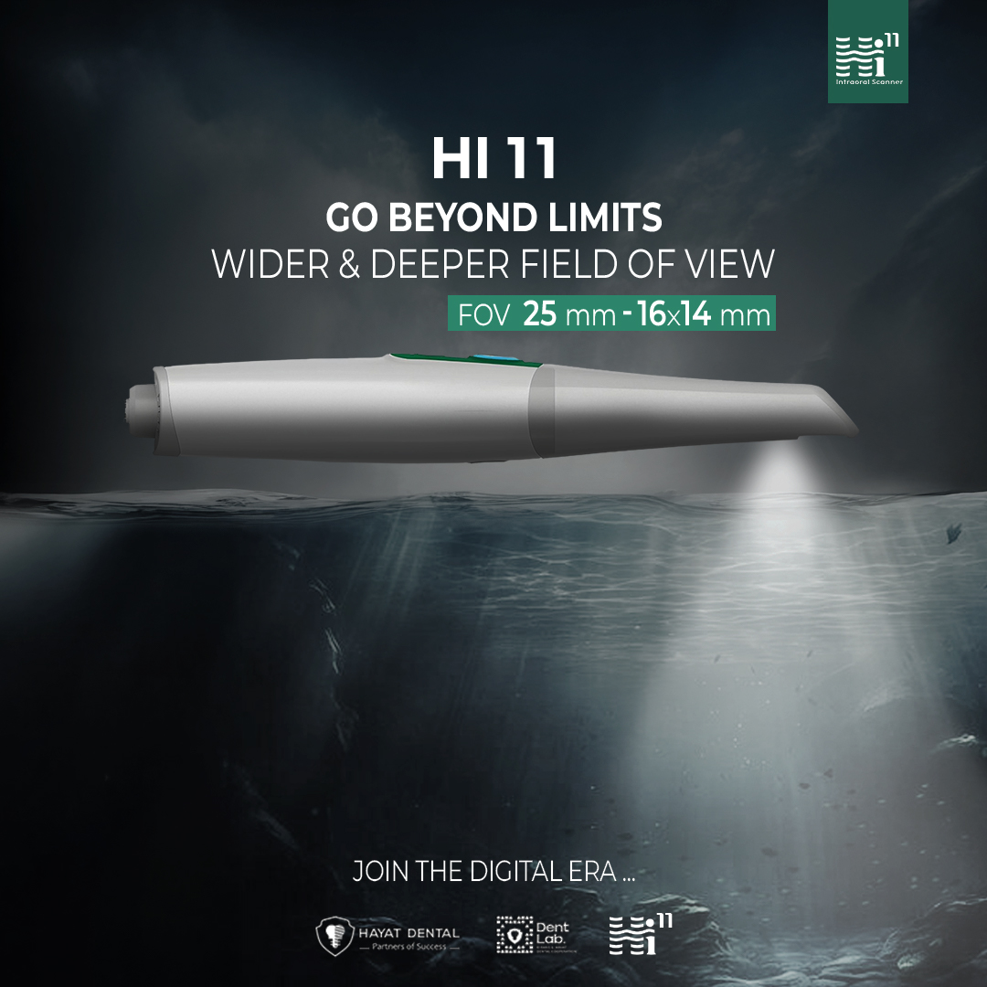
Perovskite Camera Revolutionizes Dental Imaging
Introduction: Understanding the Technology Behind Perovskite Camera
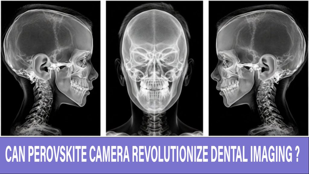
The Perovskite Camera is a groundbreaking innovation in medical and dental imaging, developed through a collaboration between Northwestern University and Soochow University. This advanced imaging system leverages perovskite crystals—materials initially recognized for their role in solar cells—to detect gamma rays with exceptional precision. These crystals are engineered to directly convert high-energy photons into electrical signals, enabling sharper and faster imaging than traditional systems.
Unlike conventional detectors that rely on scintillators and photomultiplier tubes, the Perovskite Camera uses a pixelated sensor design similar to smartphone cameras. This allows it to capture high-resolution images of bones, teeth, and soft tissues with minimal radiation exposure, setting a new benchmark for diagnostic imaging.
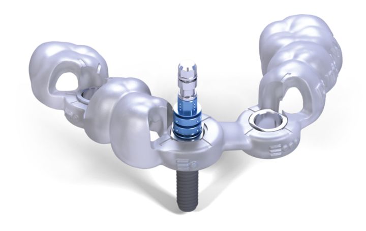
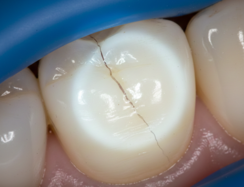
Perovskite-Camera-for-dental-imaging
Why the Perovskite Camera is a Game-Changer in Dental Imaging
Dental imaging has long relied on X-rays and cone-beam CT scans to visualize teeth, jawbones, and surrounding structures. While effective, these methods come with limitations such as radiation exposure, image noise, and restricted resolution. The advent of perovskite-based imaging introduces a new era of diagnostic clarity and safety.
With its ability to detect individual gamma-ray photons, the Perovskite Camera offers:
- Higher resolution: It can reveal fine anatomical details, including microfractures and early-stage decay.
- Lower radiation dose: Patients are exposed to significantly less radiation, making it safer for routine use.
- Improved contrast: Subtle differences in tissue density are more easily distinguishable, aiding in accurate diagnosis.
These features make it particularly valuable in dental procedures that require precise imaging, such as root canal treatments, implant planning, and orthodontic assessments.
Early Tumor Detection with Perovskite Camera Technology
One of the most promising applications of this technology is in the early detection of oral tumors. Oral cancers and jaw tumors often develop silently, with symptoms appearing only in advanced stages. Traditional imaging methods may miss small or low-density lesions, delaying diagnosis and treatment.
The Perovskite Camera’s sensitivity to gamma rays allows it to detect radiotracers like technetium-99m at extremely low doses. This enables:
- Identification of tumors at the cellular level
- Monitoring of tumor progression over time
- Guidance for minimally invasive surgical procedures
In clinical trials, the camera successfully reconstructed a 3D image of a tooth using a dose as low as 5.5 μSv—two orders of magnitude lower than standard cone-beam CT scans. This opens the door to safer and more frequent monitoring of high-risk patients.
How Perovskite Imaging Works
Perovskite crystals are grown under controlled conditions to ensure high purity and stability. When exposed to gamma rays, they generate electron-hole pairs that are read by pixelated sensors to form detailed images. This direct detection method eliminates the need for intermediate light conversion, which is common in scintillator-based systems.
Key advantages of this approach include:
- Minimized energy loss: Direct detection improves signal fidelity.
- Enhanced signal-to-noise ratio: Clearer images with less distortion.
- Real-time imaging capability: Faster diagnostics and immediate feedback during procedures.
In dental applications, this means clinicians can visualize enamel, dentin, pulp, and surrounding bone structures with unprecedented clarity.
Expanding Applications of Perovskite Camera in Dentistry
Beyond diagnostics, the Perovskite Camera has potential applications across various dental specialties:
- Implantology: Accurate bone density measurements for implant placement.
- Endodontics: Detailed visualization of root canal anatomy.
- Orthodontics: Monitoring tooth movement and bone remodeling.
- Pediatric dentistry: Safe imaging for children due to low radiation exposure.
Its compact design and cost-effective materials also make it suitable for integration into portable or intraoral devices, expanding access to advanced imaging in smaller clinics and remote areas.
Advantages of Perovskite Camera Over Traditional Imaging Systems
When comparing the Perovskite Camera to conventional imaging technologies, several advantages stand out:
- Radiation Safety: Patients receive significantly lower doses of radiation, reducing long-term health risks.
- Cost Efficiency: Perovskite materials are relatively inexpensive to produce, making the technology more affordable.
- High Sensitivity: Ability to detect low-energy gamma rays for early disease detection.
- Compact Design: Easier integration into modern dental and medical equipment.
These benefits position the Perovskite Camera as a disruptive force in the imaging industry.
The Future of Perovskite Camera in Dental Imaging
As research and development continue, the future of dental imaging looks increasingly digital, precise, and patient-friendly. Innovations on the horizon include:
- Miniaturized perovskite-based scanners: Handheld devices for chairside diagnostics.
- AI integration: Automated image analysis for faster and more accurate diagnosis.
- Tele-dentistry applications: Remote imaging and consultation capabilities.
Commercialization efforts are already underway, with companies like Actinia Inc. working to bring perovskite imaging systems to market. This could democratize access to high-quality dental care, especially in underserved regions.
Challenges and Considerations for Perovskite Camera Adoption
While the Perovskite Camera offers numerous advantages, there are challenges to address before widespread adoption:
- Material Stability: Perovskite crystals can degrade under certain environmental conditions, requiring protective coatings.
- Regulatory Approval: Medical imaging devices must meet strict safety and performance standards.
- Training Requirements: Dental professionals will need training to fully leverage the technology’s capabilities.
Addressing these challenges will be crucial for the successful integration of perovskite-based imaging into mainstream dental practice.
Conclusion: Why Perovskite Camera is the Future of Dental Imaging
The Perovskite Camera represents a paradigm shift in dental and medical imaging. By combining high-resolution imaging, low radiation exposure, and cost-effective design, it promises to enhance diagnostic accuracy and patient safety. As commercialization progresses and AI-driven features are integrated, this technology could become a standard tool in dental clinics worldwide.
For patients, this means safer, faster, and more accurate diagnoses. For dental professionals, it opens new possibilities for precision care and improved treatment outcomes. The future of imaging is here—and it’s powered by perovskite.
References:
1- First ‘Perovskite Camera’ Can See Inside the Human Body.
3- Recent Progress in Halide Perovskite Radiation Detectors for Gamma-Ray Spectroscopy.
Recent Articles


Introduction: Understanding the Technology Behind Perovskite Camera
The Perovskite Camera is a groundbreaking innovation in medical and dental imaging, developed through a collaboration between Northwestern University and Soochow University. This advanced imaging system leverages perovskite crystals—materials initially recognized for their role in solar cells—to detect gamma rays with exceptional precision. These crystals are engineered to directly convert high-energy photons into electrical signals, enabling sharper and faster imaging than traditional systems.
Unlike conventional detectors that rely on scintillators and photomultiplier tubes, the Perovskite Camera uses a pixelated sensor design similar to smartphone cameras. This allows it to capture high-resolution images of bones, teeth, and soft tissues with minimal radiation exposure, setting a new benchmark for diagnostic imaging.
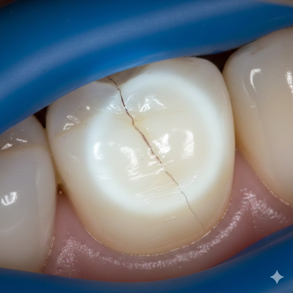
Perovskite-Camera-for-dental-imaging
Why the Perovskite Camera is a Game-Changer in Dental Imaging
Dental imaging has long relied on X-rays and cone-beam CT scans to visualize teeth, jawbones, and surrounding structures. While effective, these methods come with limitations such as radiation exposure, image noise, and restricted resolution. The advent of perovskite-based imaging introduces a new era of diagnostic clarity and safety.
With its ability to detect individual gamma-ray photons, the Perovskite Camera offers:
- Higher resolution: It can reveal fine anatomical details, including microfractures and early-stage decay.
- Lower radiation dose: Patients are exposed to significantly less radiation, making it safer for routine use.
- Improved contrast: Subtle differences in tissue density are more easily distinguishable, aiding in accurate diagnosis.
These features make it particularly valuable in dental procedures that require precise imaging, such as root canal treatments, implant planning, and orthodontic assessments.
Early Tumor Detection with Perovskite Camera Technology
One of the most promising applications of this technology is in the early detection of oral tumors. Oral cancers and jaw tumors often develop silently, with symptoms appearing only in advanced stages. Traditional imaging methods may miss small or low-density lesions, delaying diagnosis and treatment.
The Perovskite Camera’s sensitivity to gamma rays allows it to detect radiotracers like technetium-99m at extremely low doses. This enables:
- Identification of tumors at the cellular level
- Monitoring of tumor progression over time
- Guidance for minimally invasive surgical procedures
In clinical trials, the camera successfully reconstructed a 3D image of a tooth using a dose as low as 5.5 μSv—two orders of magnitude lower than standard cone-beam CT scans. This opens the door to safer and more frequent monitoring of high-risk patients.
How Perovskite Imaging Works
Perovskite crystals are grown under controlled conditions to ensure high purity and stability. When exposed to gamma rays, they generate electron-hole pairs that are read by pixelated sensors to form detailed images. This direct detection method eliminates the need for intermediate light conversion, which is common in scintillator-based systems.
Key advantages of this approach include:
- Minimized energy loss: Direct detection improves signal fidelity.
- Enhanced signal-to-noise ratio: Clearer images with less distortion.
- Real-time imaging capability: Faster diagnostics and immediate feedback during procedures.
In dental applications, this means clinicians can visualize enamel, dentin, pulp, and surrounding bone structures with unprecedented clarity.
Expanding Applications of Perovskite Camera in Dentistry
Beyond diagnostics, the Perovskite Camera has potential applications across various dental specialties:
- Implantology: Accurate bone density measurements for implant placement.
- Endodontics: Detailed visualization of root canal anatomy.
- Orthodontics: Monitoring tooth movement and bone remodeling.
- Pediatric dentistry: Safe imaging for children due to low radiation exposure.
Its compact design and cost-effective materials also make it suitable for integration into portable or intraoral devices, expanding access to advanced imaging in smaller clinics and remote areas.
Advantages of Perovskite Camera Over Traditional Imaging Systems
When comparing the Perovskite Camera to conventional imaging technologies, several advantages stand out:
- Radiation Safety: Patients receive significantly lower doses of radiation, reducing long-term health risks.
- Cost Efficiency: Perovskite materials are relatively inexpensive to produce, making the technology more affordable.
- High Sensitivity: Ability to detect low-energy gamma rays for early disease detection.
- Compact Design: Easier integration into modern dental and medical equipment.
These benefits position the Perovskite Camera as a disruptive force in the imaging industry.
The Future of Perovskite Camera in Dental Imaging
As research and development continue, the future of dental imaging looks increasingly digital, precise, and patient-friendly. Innovations on the horizon include:
- Miniaturized perovskite-based scanners: Handheld devices for chairside diagnostics.
- AI integration: Automated image analysis for faster and more accurate diagnosis.
- Tele-dentistry applications: Remote imaging and consultation capabilities.
Commercialization efforts are already underway, with companies like Actinia Inc. working to bring perovskite imaging systems to market. This could democratize access to high-quality dental care, especially in underserved regions.
Challenges and Considerations for Perovskite Camera Adoption
While the Perovskite Camera offers numerous advantages, there are challenges to address before widespread adoption:
- Material Stability: Perovskite crystals can degrade under certain environmental conditions, requiring protective coatings.
- Regulatory Approval: Medical imaging devices must meet strict safety and performance standards.
- Training Requirements: Dental professionals will need training to fully leverage the technology’s capabilities.
Addressing these challenges will be crucial for the successful integration of perovskite-based imaging into mainstream dental practice.
Conclusion: Why Perovskite Camera is the Future of Dental Imaging
The Perovskite Camera represents a paradigm shift in dental and medical imaging. By combining high-resolution imaging, low radiation exposure, and cost-effective design, it promises to enhance diagnostic accuracy and patient safety. As commercialization progresses and AI-driven features are integrated, this technology could become a standard tool in dental clinics worldwide.
For patients, this means safer, faster, and more accurate diagnoses. For dental professionals, it opens new possibilities for precision care and improved treatment outcomes. The future of imaging is here—and it’s powered by perovskite.
References:
1- First ‘Perovskite Camera’ Can See Inside the Human Body.
3- Recent Progress in Halide Perovskite Radiation Detectors for Gamma-Ray Spectroscopy.
Recent Articles
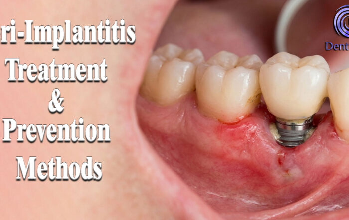
Peri-Implantitis Treatment & Prevention Methods
Peri-Implantitis Treatment & Prevention Methods Peri-implantitis remains one of the most significant biological complications affecting dental implants, posing a serious threat to long-term implant [...]
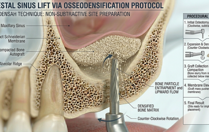
Latest Sinus Lifting Techniques in Modern Implant Dentistry
Latest Sinus Lifting Techniques in Modern Implant Dentistry A Comprehensive Clinical Review Introduction to Sinus Lifting in Implantology Sinus lifting, or maxillary sinus floor [...]
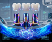
Biomechanics of the Triple Abutment & BOPiT Concept
Dental Biomechanics · Implant Science · Clinical Evidence Biomechanics of the Triple Abutment & BOPiT Concept How a saddle-shaped mathematical surface is rewriting the rules of load distribution [...]
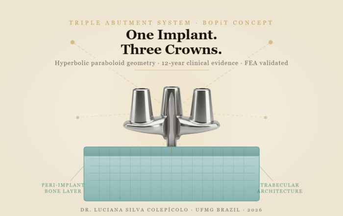
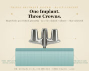
The Woman Who Proved One Implant Could Hold Three Crowns
The Woman Who Proved One Implant Could Hold Three Crowns While the dental establishment looked away, Dr. Luciana Colepícolo spent 12 years building the [...]
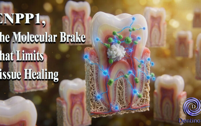
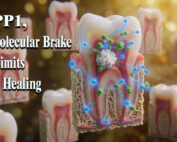
ENPP1, The Molecular Brake That Limits Tissue Healing
ENPP1, The Molecular Brake That Limits Tissue Healing Introduction: A New Biological Barrier to Dental Regeneration Meet ENPP1—a protein most dentists have never heard [...]
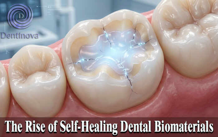
The Rise of Self‑Healing Dental Biomaterials
The Rise of Self‑Healing Dental Biomaterials Introduction In modern restorative dentistry, durability and longevity of materials remain among the greatest clinical challenges. Traditional dental [...]