

A Breakthrough for Gingival Inflammation Detection Using IOS
Transforming Dental Diagnostics with Digital Technology
Gingivitis, the earliest form of periodontal disease, is notoriously common, affecting an estimated 50–80% of adults worldwide, making the gingival inflammation detection is a must. Traditional diagnosis largely depends on visual examination and the bleeding on probing (BOP%) index, which, although useful, is subjective and depends heavily on operator skill and technique.
A groundbreaking study titled “Colorimetric analysis of intraoral scans: A novel approach for detecting gingival inflammation” introduces a new paradigm: an objective, digital, and colorimetric method that analyzes gum health through color data extracted from intraoral scans. This approach not only improves diagnostic accuracy but also paves the way for more consistent monitoring in both clinics and teledentistry settings.
Let’s explore how this innovative technique works, why it matters, and what it could mean for the future of dental care.
https://store.dentinova.co.uk/product/runyes-3ds-3-0-intraoral-scanner-ios/
Transforming Dental Diagnostics with Digital Technology
Gingivitis, the earliest form of periodontal disease, is notoriously common, affecting an estimated 50–80% of adults worldwide. Traditional diagnosis largely depends on visual examination and the bleeding on probing (BOP%) index, which, although useful, is subjective and depends heavily on operator skill and technique.
A groundbreaking study titled “Colorimetric analysis of intraoral scans: A novel approach for detecting gingival inflammation” introduces a new paradigm: an objective, digital, and colorimetric method that analyzes gum health through color data extracted from intraoral scans. This approach not only improves diagnostic accuracy but also paves the way for more consistent monitoring in both clinics and teledentistry settings.
Let’s explore how this innovative technique works, why it matters, and what it could mean for the future of dental care.
✅ The Need for a New Approach
Periodontal disease starts quietly. Subtle color changes in the gingiva—such as increased redness and swelling—often precede overt symptoms like bleeding and pain.
Current clinical standards, like bleeding on probing and plaque indices, have limitations:
Operator dependence: Accuracy varies based on the clinician’s experience.
Patient discomfort: Probing can be invasive and cause anxiety.
Limited scalability: Difficult to repeat frequently, especially in large-scale screenings or remote care.
The study aimed to overcome these limitations using colorimetric data analysis, leveraging digital technology to transform subjective visual assessments into quantifiable metrics.
📸 How Colorimetric Analysis Works: Step by Step
1. Intraoral Scanning
Researchers used intraoral scanners to capture high-resolution digital models of participants’ gums, specifically a 2 mm strip along the buccal gingival margin of the upper anterior teeth.
This region is highly visible, easily accessible, and particularly prone to inflammation, making it ideal for color analysis.
2. Image Processing
Advanced software processed these scans, focusing on color parameters:
HSV (Hue, Saturation, Value)
CIELAB color space: a*, b*, and L* components
This digital analysis extracts tiny variations in redness (a*), yellowness (b*), and saturation, which often indicate inflammation.
3. Statistical Validation
Using linear regression, the researchers correlated color metrics to bleeding on probing percentages (BOP%), the established gold standard for gingivitis detection.
Key results:
CIELAB a*: AUC ~ 70%
CIELAB b*: AUC ~ 79.5%
Color saturation: AUC ~ 80.8%
These scores place the method within the acceptable to excellent diagnostic accuracy range—impressive for a non-invasive tool.
🌍 Broader Implications for Dentistry
🔄 Continuous Monitoring
Patients could have periodic scans, even at home, to track gum health over time.
Dentists could detect deterioration before clinical signs appear.
🏥 Standardized Care
Reduces variability between practitioners and clinics, ensuring consistent diagnosis.
🌱 Prevention Focus
Early detection promotes preventive care, potentially lowering treatment costs and improving patient outcomes.
🔬 Research Catalyst
Digital colorimetric data opens new research avenues, such as studying the progression from gingivitis to periodontitis.
📈 SEO Focus: Keywords & Phrases
To help this topic rank highly on search engines, it integrates key phrases like:
Digital gingivitis detection
Intraoral scan gingival analysis
Colorimetric dental diagnostics
CIELAB gum color analysis
An objective gum inflammation tool
By weaving these phrases into content, the article appeals to both clinical audiences and search engine algorithms.
✨ The Future Outlook
While this study is pioneering, it’s also a stepping stone. Further research could:
Validate across diverse populations and different intraoral scanners.
Integrate with AI to refine accuracy.
Expand beyond gingivitis to detect early periodontitis or mucosal conditions.
The ultimate goal? Transform dental check-ups from subjective snapshots into data-driven, predictive health models.
✅ Conclusion
The colorimetric analysis of intraoral scans is more than an academic innovation—it’s a blueprint for the future of dentistry.
By translating subtle color shifts into objective, measurable data, it empowers clinicians to detect, monitor, and treat gingival inflammation earlier and more accurately.
With continued research and digital integration, this approach could become a standard tool in clinics worldwide, advancing dental care from reactive treatment to proactive prevention.
📚 Reference
Hassan, M. A., et al. (2025). Colorimetric analysis of intraoral scans: A novel approach for detecting gingival inflammation. Journal of Periodontology. DOI: 10.1002/JPER.24-0044
Recent Articles


Transforming Dental Diagnostics with Digital Technology
Gingivitis, the earliest form of periodontal disease, is notoriously common, affecting an estimated 50–80% of adults worldwide, making the gingival inflammation detection is a must. Traditional diagnosis largely depends on visual examination and the bleeding on probing (BOP%) index, which, although useful, is subjective and depends heavily on operator skill and technique.
A groundbreaking study titled “Colorimetric analysis of intraoral scans: A novel approach for detecting gingival inflammation” introduces a new paradigm: an objective, digital, and colorimetric method that analyzes gum health through color data extracted from intraoral scans. This approach not only improves diagnostic accuracy but also paves the way for more consistent monitoring in both clinics and teledentistry settings.
Let’s explore how this innovative technique works, why it matters, and what it could mean for the future of dental care.
https://store.dentinova.co.uk/product/runyes-3ds-3-0-intraoral-scanner-ios/
Transforming Dental Diagnostics with Digital Technology
Gingivitis, the earliest form of periodontal disease, is notoriously common, affecting an estimated 50–80% of adults worldwide. Traditional diagnosis largely depends on visual examination and the bleeding on probing (BOP%) index, which, although useful, is subjective and depends heavily on operator skill and technique.
A groundbreaking study titled “Colorimetric analysis of intraoral scans: A novel approach for detecting gingival inflammation” introduces a new paradigm: an objective, digital, and colorimetric method that analyzes gum health through color data extracted from intraoral scans. This approach not only improves diagnostic accuracy but also paves the way for more consistent monitoring in both clinics and teledentistry settings.
Let’s explore how this innovative technique works, why it matters, and what it could mean for the future of dental care.
✅ The Need for a New Approach
Periodontal disease starts quietly. Subtle color changes in the gingiva—such as increased redness and swelling—often precede overt symptoms like bleeding and pain.
Current clinical standards, like bleeding on probing and plaque indices, have limitations:
Operator dependence: Accuracy varies based on the clinician’s experience.
Patient discomfort: Probing can be invasive and cause anxiety.
Limited scalability: Difficult to repeat frequently, especially in large-scale screenings or remote care.
The study aimed to overcome these limitations using colorimetric data analysis, leveraging digital technology to transform subjective visual assessments into quantifiable metrics.
📸 How Colorimetric Analysis Works: Step by Step
1. Intraoral Scanning
Researchers used intraoral scanners to capture high-resolution digital models of participants’ gums, specifically a 2 mm strip along the buccal gingival margin of the upper anterior teeth.
This region is highly visible, easily accessible, and particularly prone to inflammation, making it ideal for color analysis.
2. Image Processing
Advanced software processed these scans, focusing on color parameters:
HSV (Hue, Saturation, Value)
CIELAB color space: a*, b*, and L* components
This digital analysis extracts tiny variations in redness (a*), yellowness (b*), and saturation, which often indicate inflammation.
3. Statistical Validation
Using linear regression, the researchers correlated color metrics to bleeding on probing percentages (BOP%), the established gold standard for gingivitis detection.
Key results:
CIELAB a*: AUC ~ 70%
CIELAB b*: AUC ~ 79.5%
Color saturation: AUC ~ 80.8%
These scores place the method within the acceptable to excellent diagnostic accuracy range—impressive for a non-invasive tool.
🌍 Broader Implications for Dentistry
🔄 Continuous Monitoring
Patients could have periodic scans, even at home, to track gum health over time.
Dentists could detect deterioration before clinical signs appear.
🏥 Standardized Care
Reduces variability between practitioners and clinics, ensuring consistent diagnosis.
🌱 Prevention Focus
Early detection promotes preventive care, potentially lowering treatment costs and improving patient outcomes.
🔬 Research Catalyst
Digital colorimetric data opens new research avenues, such as studying the progression from gingivitis to periodontitis.
📈 SEO Focus: Keywords & Phrases
To help this topic rank highly on search engines, it integrates key phrases like:
Digital gingivitis detection
Intraoral scan gingival analysis
Colorimetric dental diagnostics
CIELAB gum color analysis
An objective gum inflammation tool
By weaving these phrases into content, the article appeals to both clinical audiences and search engine algorithms.
✨ The Future Outlook
While this study is pioneering, it’s also a stepping stone. Further research could:
Validate across diverse populations and different intraoral scanners.
Integrate with AI to refine accuracy.
Expand beyond gingivitis to detect early periodontitis or mucosal conditions.
The ultimate goal? Transform dental check-ups from subjective snapshots into data-driven, predictive health models.
✅ Conclusion
The colorimetric analysis of intraoral scans is more than an academic innovation—it’s a blueprint for the future of dentistry.
By translating subtle color shifts into objective, measurable data, it empowers clinicians to detect, monitor, and treat gingival inflammation earlier and more accurately.
With continued research and digital integration, this approach could become a standard tool in clinics worldwide, advancing dental care from reactive treatment to proactive prevention.
📚 Reference
Hassan, M. A., et al. (2025). Colorimetric analysis of intraoral scans: A novel approach for detecting gingival inflammation. Journal of Periodontology. DOI: 10.1002/JPER.24-0044
Recent Articles
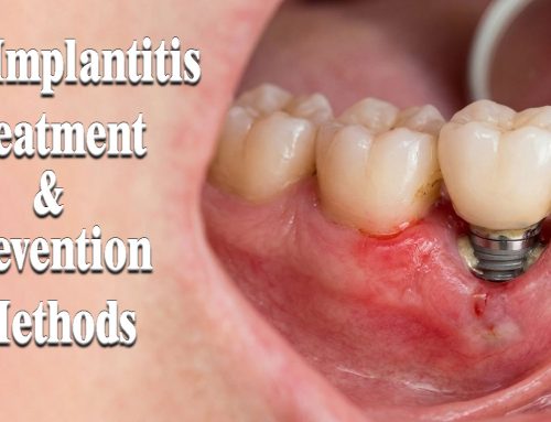
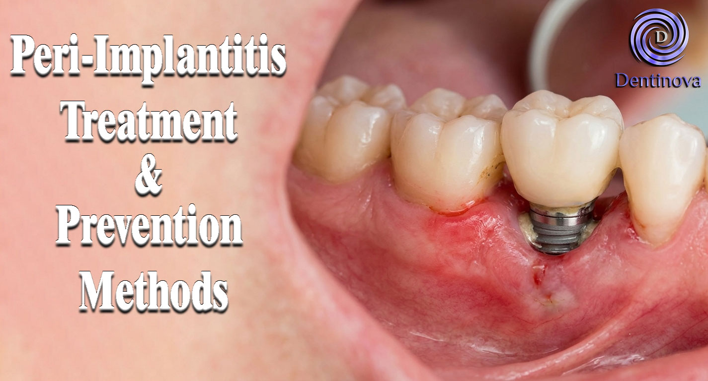
Peri-Implantitis Treatment & Prevention Methods
Peri-Implantitis Treatment & Prevention Methods Peri-implantitis remains one of the most significant biological complications affecting dental implants, posing a serious threat to long-term implant [...]
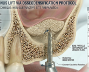
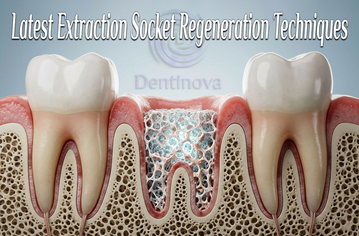
Latest Sinus Lifting Techniques in Modern Implant Dentistry
Latest Sinus Lifting Techniques in Modern Implant Dentistry A Comprehensive Clinical Review Introduction to Sinus Lifting in Implantology Sinus lifting, or maxillary sinus floor [...]
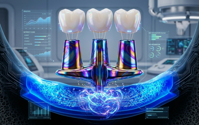
Biomechanics of the Triple Abutment & BOPiT Concept
Dental Biomechanics · Implant Science · Clinical Evidence Biomechanics of the Triple Abutment & BOPiT Concept How a saddle-shaped mathematical surface is rewriting the rules of load distribution [...]
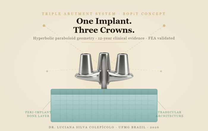
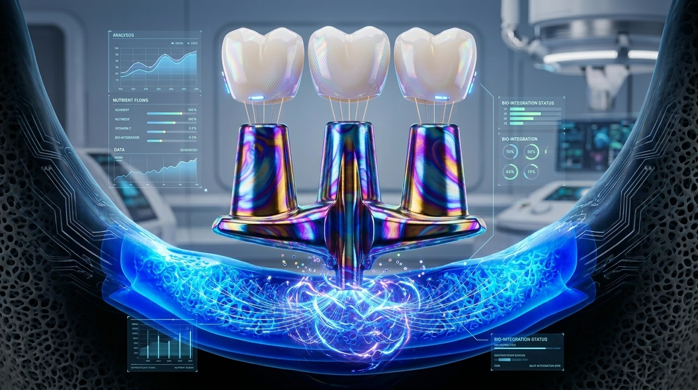
The Woman Who Proved One Implant Could Hold Three Crowns
The Woman Who Proved One Implant Could Hold Three Crowns While the dental establishment looked away, Dr. Luciana Colepícolo spent 12 years building the [...]
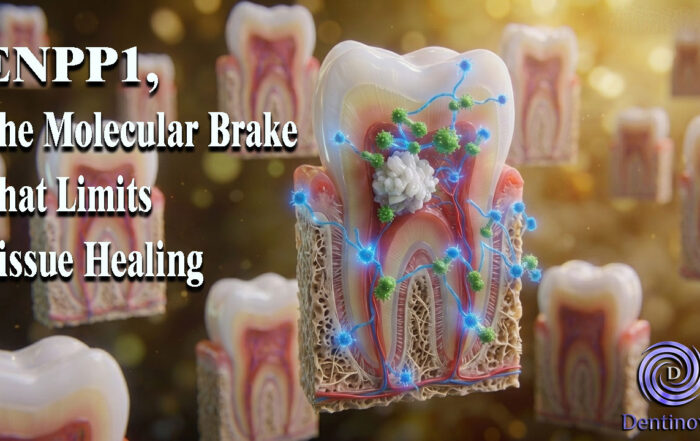
ENPP1, The Molecular Brake That Limits Tissue Healing
ENPP1, The Molecular Brake That Limits Tissue Healing Introduction: A New Biological Barrier to Dental Regeneration Meet ENPP1—a protein most dentists have never heard [...]
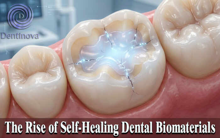
The Rise of Self‑Healing Dental Biomaterials
The Rise of Self‑Healing Dental Biomaterials Introduction In modern restorative dentistry, durability and longevity of materials remain among the greatest clinical challenges. Traditional dental [...]
