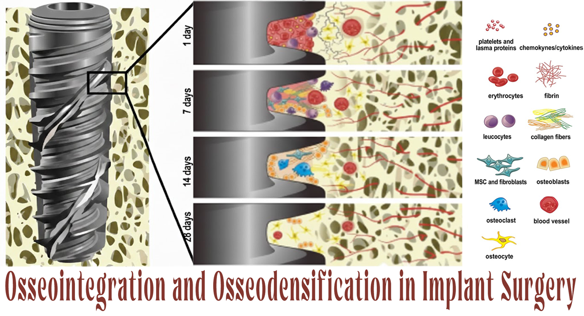
Osseointegration and Osseodensification in Implant Surgery
Introduction
Achieving predictable osseointegration remains the cornerstone of long-term dental implant success, particularly in the anterior maxilla, where esthetic demands and bone quality present unique surgical challenges. Bone density, primary stability, and atraumatic site preparation directly influence implant survival and peri-implant tissue stability. In recent years, osseodensification has emerged as a paradigm-shifting osteotomy technique designed to preserve and densify bone rather than remove it. When combined with biologic modifiers such as Advanced Platelet-Rich Fibrin (A-PRF), this approach may further enhance the biological environment for osseointegration.
This article translates current clinical evidence into a practical, surgeon-focused discussion on how osseodensification, used alone or with A-PRF, affects implant stability, bone density, and crestal bone behavior in the anterior maxilla.
Osseointegration and Primary Stability: A Biological Imperative
Osseointegration is a dynamic, biologically mediated process involving a direct structural and functional connection between living bone and the implant surface. Among the many variables influencing this process, primary stability plays a decisive role, particularly during the early healing phase. Bone density, implant macro-design, and surgical technique collectively determine the degree of micromotion at the bone–implant interface.
Conventional subtractive drilling techniques often compromise trabecular bone, especially in low-density maxillary bone. Excessive bone removal, elliptical osteotomies, and thermal trauma can prolong remodeling and reduce early stability. These limitations have driven the adoption of alternative preparation methods aimed at enhancing bone preservation and mechanical engagement at placement.
Osseodensification and Osseointegration in the Anterior Maxilla
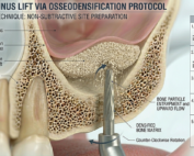
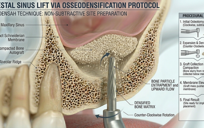
Osseodensification is a non-subtractive osteotomy technique performed using specially designed burs operating in a counter-clockwise, densifying mode. Instead of excavating bone, these burs compact trabecular bone laterally and apically, increasing peri-implant bone density and improving implant–bone contact.
In the anterior maxilla, where bone is frequently classified as D3 or D4, osseodensification offers several advantages:
Increased insertion torque and primary stability
Autografting of compacted bone particles into the osteotomy walls
Improved ridge expansion without additional grafting
Reduced microfractures and thermal insult
These biomechanical and biological effects create a more favorable environment for predictable osseointegration in esthetically critical regions.
The Role of A-PRF in Enhancing Osseointegration
Advanced Platelet-Rich Fibrin (A-PRF) represents an evolution of autologous platelet concentrates. Prepared using lower centrifugation forces, A-PRF contains higher concentrations of leukocytes and growth factors, including PDGF, VEGF, TGF-β, and IGF. These bioactive molecules are gradually released over time, supporting angiogenesis, osteogenesis, and soft-tissue healing.
When introduced into an osseodensified osteotomy, A-PRF acts as a biologic scaffold that complements mechanical bone compaction. The synergistic effect promotes faster bone maturation and may positively influence early implant stability and secondary osseointegration.
Osseodensification and Implant Stability: Clinical Evidence
Clinical evaluation of implant stability using resonance frequency analysis (ISQ values) demonstrates a consistent advantage when osseodensification is combined with A-PRF. Implants placed with this combined approach show significantly higher primary stability at placement and superior secondary stability after six months compared to osseodensification alone.
This improvement can be attributed to:
Enhanced bone-implant contact from densified trabecular bone
Accelerated bone regeneration mediated by A-PRF growth factors
Improved mechanical interlocking during early healing
Higher ISQ values at both time points suggest a more rapid transition from mechanical to biological stability, a critical factor in esthetic zone implants.
Radiographic Outcomes: Bone Density and Crestal Bone Preservation
Radiographic assessment using CBCT provides objective insight into peri-implant bone behavior. Bone density measurements reveal a statistically significant increase after six months when A-PRF is used in conjunction with osseodensification. This finding supports the concept that biologically active fibrin matrices enhance bone remodeling and maturation beyond what mechanical densification alone can achieve.
In contrast, crestal bone levels remain stable and comparable between both approaches. The absence of significant marginal bone loss indicates that osseodensification, whether used alone or with A-PRF, does not negatively affect crestal bone preservation in the short term. This is clinically relevant, as marginal bone stability is closely tied to esthetic outcomes and long-term implant success.
Clinical Implications for Implant Surgeons
From a practical standpoint, combining osseodensification with A-PRF offers several advantages for implant placement in the anterior maxilla:
Improved primary and secondary implant stability
Increased peri-implant bone density without additional grafting
Favorable soft-tissue healing and reduced postoperative morbidity
Predictable outcomes in low-density bone
Importantly, postoperative pain, edema, and complication rates appear comparable between both techniques, suggesting that the addition of A-PRF does not increase patient morbidity. Faster wound healing and improved tissue response may further enhance patient satisfaction and clinician confidence.
Best Osseodensification Tools in the Market
Densah® Burs – Versah
The Densah® Bur system by Versah is widely regarded as the gold standard in osseodensification tools and is one of the earliest and most thoroughly validated technologies designed specifically for osteotomy preparation with densification in mind. These burs are engineered with a proprietary multi-flute design that allows them to work in two distinct drilling modes: cutting in a clockwise direction and densifying in a counter-clockwise direction. In densifying mode, the burs compact bone laterally and apically, increasing peri-implant bone density while minimizing bone removal. This unique capability makes the Densah® burs particularly effective in soft bone conditions like those often encountered in the maxilla, where enhanced primary stability is critical. With compatibility across a wide range of implant diameters and surgical indications, including ridge expansion and crestal sinus lift, the Versah Densah® system supports predictable osseointegration and has become a mainstay for clinicians embracing modern implant site preparation techniques.
Universal Osseodensification Bur Kits
Universal osseodensification bur kits represent comprehensive systems designed to support a broad range of implant diameters with interchangeable bur sizes. These kits typically include multiple tapered osseodensification burs, pilot drills, parallel pins, and depth markers, permitting precise preparation in both cutting and densifying modes. By offering a wide selection of sizes, from 2.0 mm through 5.5 mm, these kits allow clinicians to tailor the osteotomy to the specific anatomical needs of each patient. Their external irrigation design helps manage heat generation, reduce surgical trauma, and enhance bone preservation. Universal kits are typically more affordable and may be ideal for practices that require flexibility across multiple implant systems. Their versatility makes them valuable assets for clinicians seeking predictable osseointegration and improved primary stability without investing in brand-specific systems.
Dental Osseodensification Bur Kits (Multi-Piece Sets)
Multi-piece dental osseodensification bur kits, such as 21-, 24-, or 29-piece sets, offer extensive instrumentation for implant site preparation and bone preservation. These kits provide an array of bur sizes, depth stoppers, and specialized instruments that facilitate precise bone compaction and density enhancement. Designed with surgical-grade materials and autoclavable components, these sets emphasize durability and repeated use in clinical settings. Dental professionals benefit from the inclusivity of detailed bur diameters and stoppers that support controlled penetration depths, reducing procedural variability and improving surgical confidence. Such kits are particularly effective when managing diverse clinical scenarios, including narrow ridges, compromised bone volume, or simultaneous ridge expansion. By integrating a comprehensive bur selection with consistent performance characteristics, multi-piece osseodensification kits help clinicians achieve predictable osseointegration and long-term implant success.
Conclusion
Osseodensification represents a biologically respectful and mechanically advantageous approach to implant site preparation, particularly in the anterior maxilla. When combined with Advanced Platelet-Rich Fibrin, this technique demonstrates measurable improvements in implant stability and bone density without compromising crestal bone levels. For implant surgeons seeking predictable osseointegration in esthetically demanding cases, this combined strategy offers a safe, effective, and evidence-based solution.
Reference:
Mohamed M. Elkamah* BDS, Magda M. Saleh2 PhD, Saeeda M. Osman2 PhD, Mona S. Oraby3 PhD
Frequently Asked Questions (FAQ)
What is the main advantage of osseodensification over conventional drilling?
Osseodensification preserves and compacts bone rather than removing it, leading to increased bone density, improved primary stability, and enhanced osseointegration, especially in low-density bone.
Does adding A-PRF significantly improve implant outcomes?
Yes. Clinical evidence shows that A-PRF enhances secondary stability and peri-implant bone density by promoting angiogenesis and osteogenesis during healing.
Is osseodensification suitable for the anterior maxilla?
Absolutely. It is particularly beneficial in the anterior maxilla due to frequent low bone density and high esthetic demands.
Does A-PRF reduce postoperative pain or swelling?
While pain and edema are similar with or without A-PRF, faster soft-tissue healing and improved wound quality are commonly observed.
Can osseodensification eliminate the need for bone grafting?
In selected cases with adequate ridge width, osseodensification may reduce or eliminate the need for additional grafting by expanding and densifying existing bone.
Is this technique compatible with immediate or early loading protocols?
Improved primary and secondary stability suggests potential suitability, but loading decisions should be based on individual clinical assessment and ISQ values.
Recent Articles


















Introduction
Achieving predictable osseointegration remains the cornerstone of long-term dental implant success, particularly in the anterior maxilla, where esthetic demands and bone quality present unique surgical challenges. Bone density, primary stability, and atraumatic site preparation directly influence implant survival and peri-implant tissue stability. In recent years, osseodensification has emerged as a paradigm-shifting osteotomy technique designed to preserve and densify bone rather than remove it. When combined with biologic modifiers such as Advanced Platelet-Rich Fibrin (A-PRF), this approach may further enhance the biological environment for osseointegration.
This article translates current clinical evidence into a practical, surgeon-focused discussion on how osseodensification, used alone or with A-PRF, affects implant stability, bone density, and crestal bone behavior in the anterior maxilla.
Osseointegration and Primary Stability: A Biological Imperative
Osseointegration is a dynamic, biologically mediated process involving a direct structural and functional connection between living bone and the implant surface. Among the many variables influencing this process, primary stability plays a decisive role, particularly during the early healing phase. Bone density, implant macro-design, and surgical technique collectively determine the degree of micromotion at the bone–implant interface.
Conventional subtractive drilling techniques often compromise trabecular bone, especially in low-density maxillary bone. Excessive bone removal, elliptical osteotomies, and thermal trauma can prolong remodeling and reduce early stability. These limitations have driven the adoption of alternative preparation methods aimed at enhancing bone preservation and mechanical engagement at placement.
Osseodensification and Osseointegration in the Anterior Maxilla
Osseodensification is a non-subtractive osteotomy technique performed using specially designed burs operating in a counter-clockwise, densifying mode. Instead of excavating bone, these burs compact trabecular bone laterally and apically, increasing peri-implant bone density and improving implant–bone contact.
In the anterior maxilla, where bone is frequently classified as D3 or D4, osseodensification offers several advantages:
Increased insertion torque and primary stability
Autografting of compacted bone particles into the osteotomy walls
Improved ridge expansion without additional grafting
Reduced microfractures and thermal insult
These biomechanical and biological effects create a more favorable environment for predictable osseointegration in esthetically critical regions.
The Role of A-PRF in Enhancing Osseointegration
Advanced Platelet-Rich Fibrin (A-PRF) represents an evolution of autologous platelet concentrates. Prepared using lower centrifugation forces, A-PRF contains higher concentrations of leukocytes and growth factors, including PDGF, VEGF, TGF-β, and IGF. These bioactive molecules are gradually released over time, supporting angiogenesis, osteogenesis, and soft-tissue healing.
When introduced into an osseodensified osteotomy, A-PRF acts as a biologic scaffold that complements mechanical bone compaction. The synergistic effect promotes faster bone maturation and may positively influence early implant stability and secondary osseointegration.
Osseodensification and Implant Stability: Clinical Evidence
Clinical evaluation of implant stability using resonance frequency analysis (ISQ values) demonstrates a consistent advantage when osseodensification is combined with A-PRF. Implants placed with this combined approach show significantly higher primary stability at placement and superior secondary stability after six months compared to osseodensification alone.
This improvement can be attributed to:
Enhanced bone-implant contact from densified trabecular bone
Accelerated bone regeneration mediated by A-PRF growth factors
Improved mechanical interlocking during early healing
Higher ISQ values at both time points suggest a more rapid transition from mechanical to biological stability, a critical factor in esthetic zone implants.
Radiographic Outcomes: Bone Density and Crestal Bone Preservation
Radiographic assessment using CBCT provides objective insight into peri-implant bone behavior. Bone density measurements reveal a statistically significant increase after six months when A-PRF is used in conjunction with osseodensification. This finding supports the concept that biologically active fibrin matrices enhance bone remodeling and maturation beyond what mechanical densification alone can achieve.
In contrast, crestal bone levels remain stable and comparable between both approaches. The absence of significant marginal bone loss indicates that osseodensification, whether used alone or with A-PRF, does not negatively affect crestal bone preservation in the short term. This is clinically relevant, as marginal bone stability is closely tied to esthetic outcomes and long-term implant success.
Clinical Implications for Implant Surgeons
From a practical standpoint, combining osseodensification with A-PRF offers several advantages for implant placement in the anterior maxilla:
Improved primary and secondary implant stability
Increased peri-implant bone density without additional grafting
Favorable soft-tissue healing and reduced postoperative morbidity
Predictable outcomes in low-density bone
Importantly, postoperative pain, edema, and complication rates appear comparable between both techniques, suggesting that the addition of A-PRF does not increase patient morbidity. Faster wound healing and improved tissue response may further enhance patient satisfaction and clinician confidence.
Best Osseodensification Tools in the Market
Densah® Burs – Versah
The Densah® Bur system by Versah is widely regarded as the gold standard in osseodensification tools and is one of the earliest and most thoroughly validated technologies designed specifically for osteotomy preparation with densification in mind. These burs are engineered with a proprietary multi-flute design that allows them to work in two distinct drilling modes: cutting in a clockwise direction and densifying in a counter-clockwise direction. In densifying mode, the burs compact bone laterally and apically, increasing peri-implant bone density while minimizing bone removal. This unique capability makes the Densah® burs particularly effective in soft bone conditions like those often encountered in the maxilla, where enhanced primary stability is critical. With compatibility across a wide range of implant diameters and surgical indications, including ridge expansion and crestal sinus lift, the Versah Densah® system supports predictable osseointegration and has become a mainstay for clinicians embracing modern implant site preparation techniques.
Universal Osseodensification Bur Kits
Universal osseodensification bur kits represent comprehensive systems designed to support a broad range of implant diameters with interchangeable bur sizes. These kits typically include multiple tapered osseodensification burs, pilot drills, parallel pins, and depth markers, permitting precise preparation in both cutting and densifying modes. By offering a wide selection of sizes, from 2.0 mm through 5.5 mm, these kits allow clinicians to tailor the osteotomy to the specific anatomical needs of each patient. Their external irrigation design helps manage heat generation, reduce surgical trauma, and enhance bone preservation. Universal kits are typically more affordable and may be ideal for practices that require flexibility across multiple implant systems. Their versatility makes them valuable assets for clinicians seeking predictable osseointegration and improved primary stability without investing in brand-specific systems.
Dental Osseodensification Bur Kits (Multi-Piece Sets)
Multi-piece dental osseodensification bur kits, such as 21-, 24-, or 29-piece sets, offer extensive instrumentation for implant site preparation and bone preservation. These kits provide an array of bur sizes, depth stoppers, and specialized instruments that facilitate precise bone compaction and density enhancement. Designed with surgical-grade materials and autoclavable components, these sets emphasize durability and repeated use in clinical settings. Dental professionals benefit from the inclusivity of detailed bur diameters and stoppers that support controlled penetration depths, reducing procedural variability and improving surgical confidence. Such kits are particularly effective when managing diverse clinical scenarios, including narrow ridges, compromised bone volume, or simultaneous ridge expansion. By integrating a comprehensive bur selection with consistent performance characteristics, multi-piece osseodensification kits help clinicians achieve predictable osseointegration and long-term implant success.
Conclusion
Osseodensification represents a biologically respectful and mechanically advantageous approach to implant site preparation, particularly in the anterior maxilla. When combined with Advanced Platelet-Rich Fibrin, this technique demonstrates measurable improvements in implant stability and bone density without compromising crestal bone levels. For implant surgeons seeking predictable osseointegration in esthetically demanding cases, this combined strategy offers a safe, effective, and evidence-based solution.
Reference:
Mohamed M. Elkamah* BDS, Magda M. Saleh2 PhD, Saeeda M. Osman2 PhD, Mona S. Oraby3 PhD
Frequently Asked Questions (FAQ)
What is the main advantage of osseodensification over conventional drilling?
Osseodensification preserves and compacts bone rather than removing it, leading to increased bone density, improved primary stability, and enhanced osseointegration, especially in low-density bone.
Does adding A-PRF significantly improve implant outcomes?
Yes. Clinical evidence shows that A-PRF enhances secondary stability and peri-implant bone density by promoting angiogenesis and osteogenesis during healing.
Is osseodensification suitable for the anterior maxilla?
Absolutely. It is particularly beneficial in the anterior maxilla due to frequent low bone density and high esthetic demands.
Does A-PRF reduce postoperative pain or swelling?
While pain and edema are similar with or without A-PRF, faster soft-tissue healing and improved wound quality are commonly observed.
Can osseodensification eliminate the need for bone grafting?
In selected cases with adequate ridge width, osseodensification may reduce or eliminate the need for additional grafting by expanding and densifying existing bone.
Is this technique compatible with immediate or early loading protocols?
Improved primary and secondary stability suggests potential suitability, but loading decisions should be based on individual clinical assessment and ISQ values.
Recent Articles
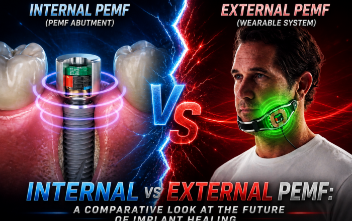
Pulsed Electromagnetic Field (PEMF) Use in Implant Dentistry
Pulsed Electromagnetic Field (PEMF) Use in Implant Dentistry Abstract and Historical Background Pulsed Electromagnetic Field (PEMF) therapy refers to the application of low-frequency electromagnetic [...]
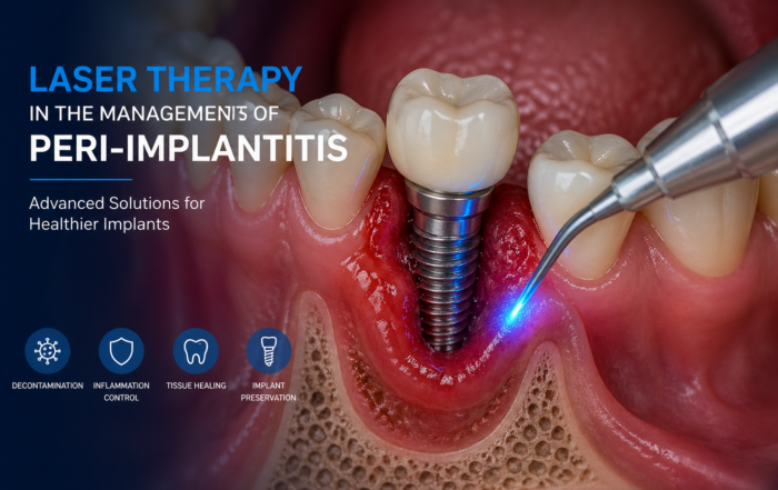
Laser Therapy in the Management of Peri-Implantitis
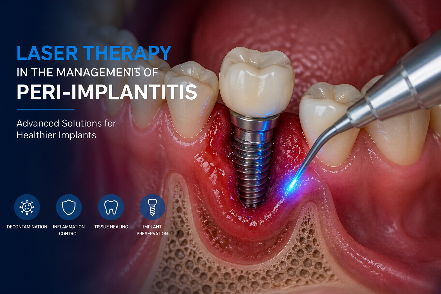
Laser Therapy in the Management of Peri-Implantitis Laser technology has become an increasingly valuable adjunct in the treatment of peri-implant diseases, particularly peri-implantitis. Its [...]
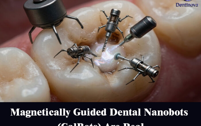
Magnetically Guided Dental Nanobots (CalBots) Are Real
Magnetically Guided Dental Nanobots (CalBots) Are Real The concept of nanobots in dentistry has long been associated with futuristic speculation. However, recent peer-reviewed research [...]
Peri-Implantitis Treatment & Prevention Methods
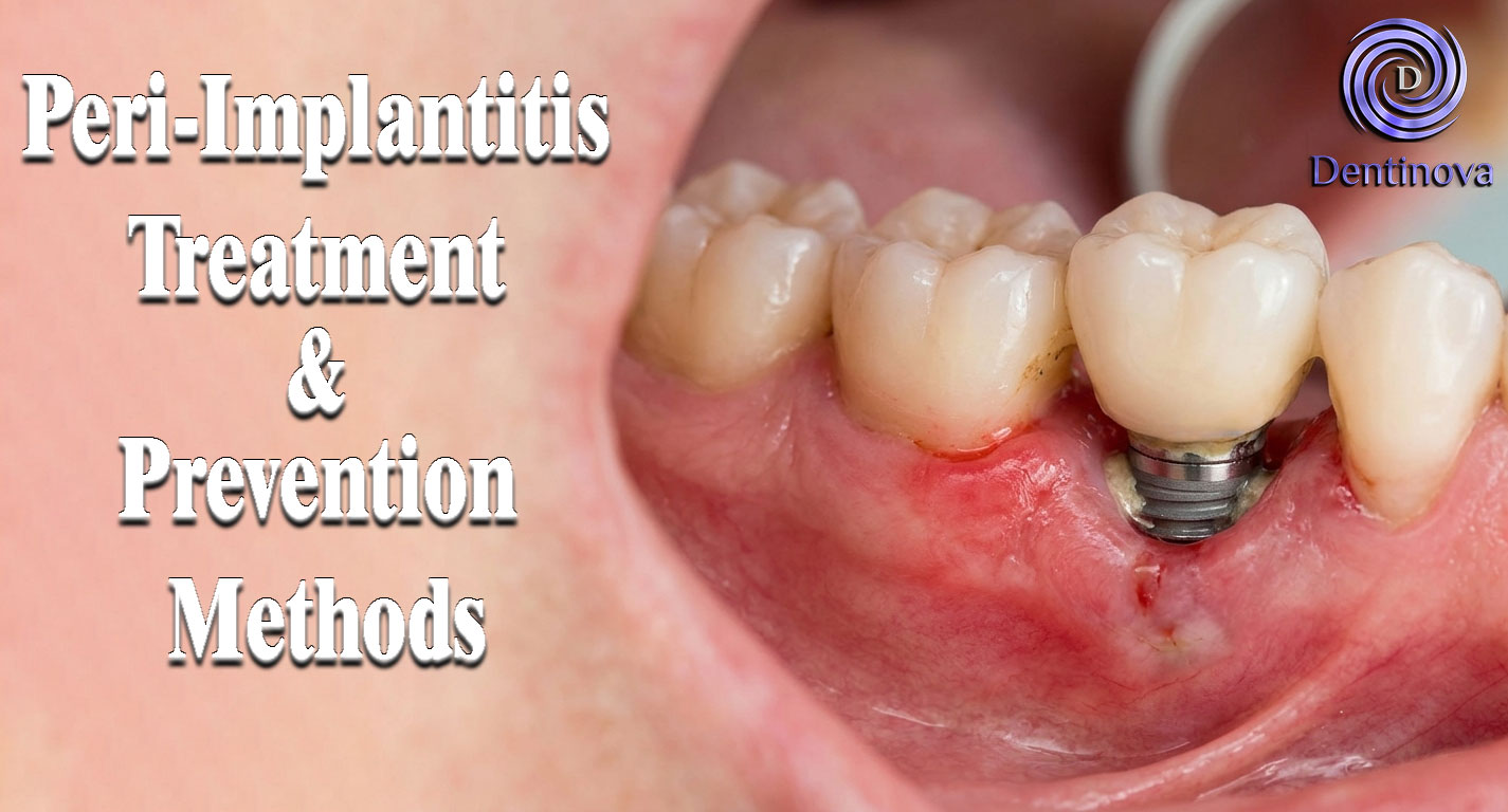
Peri-Implantitis Treatment & Prevention Methods Peri-implantitis remains one of the most significant biological complications affecting dental implants, posing a serious threat to long-term implant [...]
Latest Sinus Lifting Techniques in Modern Implant Dentistry
Latest Sinus Lifting Techniques in Modern Implant Dentistry A Comprehensive Clinical Review Introduction to Sinus Lifting in Implantology Sinus lifting, or maxillary sinus floor [...]
Biomechanics of the Triple Abutment & BOPiT Concept
Dental Biomechanics · Implant Science · Clinical Evidence Biomechanics of the Triple Abutment & BOPiT Concept How a saddle-shaped mathematical surface is rewriting the rules of load distribution [...]